
tóng-àn:Ebola virus em.png
Choân kái-sek-tō͘ (2,043 × 2,887 siōng-sò͘ , tóng-àn chiàm-liōng: 2.55 MB, MIME luī-hêng: image/png)
|
|
Che sī tùi Wikimedia Commons ín--lâi ê chi̍t hūn tóng-àn. I tī hia ê kì-su̍t-ia̍h téng-bīn ê chu-sìn hián-sī ùi ē-té. |
| Soat-bêng |
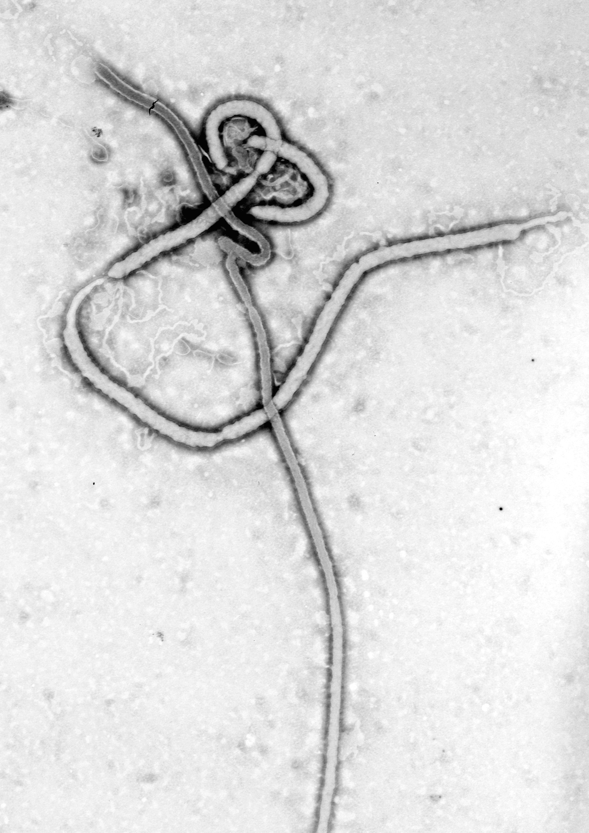
English: An electron micrograph of an Ebola viral particle showing the characteristic filamentous structure of a Filoviridae. The viral filaments can appear in images in various shapes including a 'u', '6', a coil, or branched resulting in pleomorphic particles. The filaments are reported to be between 60-80 nm in diameter, the length of a filament associated with an individual viral partial is extremely variable with Ebola particles of up to 14,000 nm in length being reported. An average length, which may represent the most infectious particles is in the region of 1000 nm.
The first electronmicrograph of Ebola was 13 October 1976 by Dr. F.A. Murphy, now at UC Davis, who was then working at the CDC. The nucleocapsid structure consists of a central channel, 20-30nm in diameter, surrounded by helically wound capsid with a diameter of 40-50nm and an interval of 5nm. 7nm glycoprotein spikes spaced 10 nm apart from each other are present within the outer envelope of the virus which is derived from the host cell membrane. Each viral particle contains one molecule of single-stranded, negative-sense RNA, which encodes the seven viral proteins.Polski: Grafika przedstawiająca wirus Ebola pochodząca z mikroskopu elektronowego ukazująca
charakterystyczną filamentową budowę tego Filowirusa. Te wirusowe filamenty występują na obrzach w różnych postaciach między innymi: "u" '6' albo sprężyny. Filamenty występują w rozmiarze 60-80 nm średnicy. Długość filamentu jest bardzo zmienna i zależna od konkretnej "cząsteczki" wirusa, w przypadku Ebola pojawiają się mające długość nawet 14,000 nm. Średnia długość, która może reprezentować wersję najbardziej zaraźliwą ma długość około 1000nm. Pierwsze zdjęcie wirusa Ebola zostało wykonane 13 Października 1976 roku przez Dr. F.A Murphy, teraz pracującego na uniwersytecie w Davis, wtedy w CDC.中文:
Source: CDC
|
|||
| Ji̍t-kî | ||||
| Chhut-chhù |
|
|||
| Chok-chiá | CDC/ Dr. Frederick A. Murphy | |||
| 授權允准 (Bô siŏh-huòi sāi ciā ùng-giông) |
|
Tóng-àn le̍k-sú
Chhi̍h ji̍t-kî/sî-kan, khoàⁿ hit sî-chūn--ê tóng-àn.
| Ji̍t-kî/Sî-kan | 細張圖 | 寸尺 | Iōng-chiá | Chù-kái | |
|---|---|---|---|---|---|
| hiān-chāi | 2014-nî 7-goe̍h 28-ji̍t (pài-it) 09:33 | | 2,043 × 2,887(2.55 MB) | Splintercellguy | Upload higher-resolution version |
| 2005-nî 5-goe̍h 24-ji̍t (pài-jī) 11:22 |  | 150 × 227(19 KB) | Knutux | An electron micrograph of an Ebola viral particle showing the characteristic filamentous structure of a Filoviridae. The viral filaments can appear in images in various shapes including a 'u', '6', a coil, or branched resul |
Iáⁿ-siōng liân-kiat
Í-hā ê ia̍h liân kàu chit ê iáⁿ-siōng:
tóng-àn hō͘ lâng sái--ê chōng-hóng
Ē-kha--ê kî-thaⁿ wiki ēng tio̍h chit--ê tóng-àn:
- af.wikipedia.org hō͘ lâng ēng--ê chêng-hêng
- als.wikipedia.org hō͘ lâng ēng--ê chêng-hêng
- ar.wikipedia.org hō͘ lâng ēng--ê chêng-hêng
- arz.wikipedia.org hō͘ lâng ēng--ê chêng-hêng
- ast.wikipedia.org hō͘ lâng ēng--ê chêng-hêng
- bn.wikipedia.org hō͘ lâng ēng--ê chêng-hêng
- ca.wikipedia.org hō͘ lâng ēng--ê chêng-hêng
- ca.wikinews.org hō͘ lâng ēng--ê chêng-hêng
- csb.wikipedia.org hō͘ lâng ēng--ê chêng-hêng
- da.wikipedia.org hō͘ lâng ēng--ê chêng-hêng
- de.wikipedia.org hō͘ lâng ēng--ê chêng-hêng
- el.wikipedia.org hō͘ lâng ēng--ê chêng-hêng
- en.wikipedia.org hō͘ lâng ēng--ê chêng-hêng
- Wikipedia:Main Page/French
- Wikipedia:WikiProject Viruses/Templates
- Virus
- Wikipedia:Top 25 Report/July 27 to August 2, 2014
- Wikipedia:Top 25 Report/August 3 to 9, 2014
- RVSV-ZEBOV vaccine
- CAd3-ZEBOV
- 2016 in science
- Wikipedia:Top 25 Report/Records
- User:M1Abramstanbks/sandbox
- Wikipedia:In the news/Posted/May 2004
- en.wikinews.org hō͘ lâng ēng--ê chêng-hêng
- eo.wikipedia.org hō͘ lâng ēng--ê chêng-hêng
- eo.wikinews.org hō͘ lâng ēng--ê chêng-hêng
- es.wikipedia.org hō͘ lâng ēng--ê chêng-hêng
- et.wikipedia.org hō͘ lâng ēng--ê chêng-hêng
- eu.wikipedia.org hō͘ lâng ēng--ê chêng-hêng
- fi.wikiversity.org hō͘ lâng ēng--ê chêng-hêng
- fr.wikipedia.org hō͘ lâng ēng--ê chêng-hêng
- fr.wikibooks.org hō͘ lâng ēng--ê chêng-hêng
- ga.wikipedia.org hō͘ lâng ēng--ê chêng-hêng
- gl.wikipedia.org hō͘ lâng ēng--ê chêng-hêng
檢視此檔案的更多全域使用狀況。