Pûi-tōa sè-pau
| Puî-tuā sè-pau (mast cell) | |
|---|---|
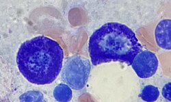
 Ū nn̄g-ê puî-tuā sè-pau teh kut-tshué lāi-té. | |
| Siông-sè | |
| System | Bián-i̍k hē-thóng |
| Sek-pia̍t hû-hō | |
| Latin-gí | mastocytus |
| TH | H2.00.03.0.01010 |
| FMA | 66784 |
Puî-tuā sè-pau (hàn-gú: 肥大細胞; ing-gú: mast cell (iah kiò-tsò mastocyte hi̍k-tsiá labrocyte[1])) sī kiat-tè tsoo-tsit (結締組織) ê siông-tsù (常駐) sè-pau, hâm-iú tsiânn-tsē hù-hâm tsoo-an hām kuann-sòo ê lia̍p-á. Kū-thé lâi-kóng, puî-tuā sè-pau sī tsi̍t-tsióng guân-tsū kut-tshué kàn sè-pau ê lia̍p sè-pau, sī bián-i̍k hām sîn-king bián-i̍k hē-thóngs ê tsi̍t-pōo-hūn. Puî-tuā sè-pau iû Paul Ehrlich tī 1877-nî huat-hiān.[2] Sui-bóng Puî-tuā sè-pau in i teh kuè-bín hām kuè-bín huán-ìng tang-tiong ê tsok-iōng jî-lâi tshut-miâ; m̄-ko puî-tuā sè-pau iah huat-hui tio̍h tiōng-iàu ê pó-hōo tsok-iōng, bi̍t-tshiat tsham-ú siong-kháu kuè-phuê, hiat-kuán senn-sîng, bián-i̍k nāi-siū, pēnn-guân-thé hông-gū kap náu tsíng-liû tang-tiong ê hiat-kuán thong-thàu sìng.[3][4]
Puî-tuā sè-pau ê guā-kuan hām kong-lîng kah līng-guā tsi̍t-tsióng pe̍h sè-pau, tsik-sī sī kinn-sìng lia̍p sè-pau beh-tsiânn sio-kâng. Sui-bóng Puî-tuā sè-pau pat hông jīn-uî sī tsū-liû ê sī kinn-sìng lia̍p sè-pau; m̄-ko sū-si̍t tsìng-bîng, tsit nn̄g-tsióng sè-pau sī iû bô kâng-khuán ê tsō-hiat phóo-hē huat-io̍k jî-lâi; in-tshú bô khó-lîng sī sio-kâng ê sè-pau.[5]
Tsù-kái[siu-kái | kái goân-sí-bé]
- ↑ "labrocytes". Memidex. goân-loē-iông tī 6 November 2018 hőng khó͘-pih. 19 February 2011 khòaⁿ--ê.
- ↑ Ehrlich, Paul (1878). "Beiträge zur Theorie und Praxis der Histologischen Färbung". Leipzig University.
- ↑ da Silva EZ, Jamur MC, Oliver C (2014). "Mast cell function: a new vision of an old cell". J. Histochem. Cytochem. 62 (10): 698–738. doi:10.1369/0022155414545334. PMC 4230976
 . PMID 25062998.
 Mast cells can recognize pathogens through different mechanisms including direct binding of pathogens or their components to PAMP receptors on the mast cell surface, binding of antibody or complement-coated bacteria to complement or immunoglobulin receptors, or recognition of endogenous peptides produced by infected or injured cells (Hofmann and Abraham 2009). The pattern of expression of these receptors varies considerably among different mast cell subtypes. TLRs (1–7 and 9), NLRs, RLRs, and receptors for complement are accountable for most mast cell innate responses
- ↑ Polyzoidis S, Koletsa T, Panagiotidou S, Ashkan K, Theoharides TC (2015). "Mast cells in meningiomas and brain inflammation". J Neuroinflammation. 12 (1): 170. doi:10.1186/s12974-015-0388-3. PMC 4573939
 . PMID 26377554.
 MCs originate from a bone marrow progenitor and subsequently develop different phenotype characteristics locally in tissues. Their range of functions is wide and includes participation in allergic reactions, innate and adaptive immunity, inflammation, and autoimmunity [34]. In the human brain, MCs can be located in various areas, such as the pituitary stalk, the pineal gland, the area postrema, the choroid plexus, thalamus, hypothalamus, and the median eminence [35]. In the meninges, they are found within the dural layer in association with vessels and terminals of meningeal nociceptors [36]. MCs have a distinct feature compared to other hematopoietic cells in that they reside in the brain [37]. MCs contain numerous granules and secrete an abundance of prestored mediators such as corticotropin-releasing hormone (CRH), neurotensin (NT), substance P (SP), tryptase, chymase, vasoactive intestinal peptide (VIP), vascular endothelial growth factor (VEGF), TNF, prostaglandins, leukotrienes, and varieties of chemokines and cytokines some of which are known to disrupt the integrity of the blood-brain barrier (BBB) [38–40].
[The] key role of MCs in inflammation [34] and in the disruption of the BBB [41–43] suggests areas of importance for novel therapy research. Increasing evidence also indicates that MCs participate in neuroinflammation directly [44–46] and through microglia stimulation [47], contributing to the pathogenesis of such conditions such as headaches, [48] autism [49], and chronic fatigue syndrome [50]. In fact, a recent review indicated that peripheral inflammatory stimuli can cause microglia activation [51], thus possibly involving MCs outside the brain. - ↑ Franco CB, Chen CC, Drukker M, Weissman IL, Galli SJ (2010). "Distinguishing mast cell and granulocyte differentiation at the single-cell level". Cell Stem Cell. 6 (4): 361–8. doi:10.1016/j.stem.2010.02.013. PMC 2852254
 . PMID 20362540.
Tsham -ua̍t[siu-kái | kái goân-sí-bé]
- Allergy
- Mast cell activation syndrome
- Diamine oxidase
- Granulocyte
- Food intolerance
- Histamine
- Histamine intolerance
- Histamine N-methyltransferase or HNMT
- List of distinct cell types in the adult human body
Guā-pōo liân-kiat[siu-kái | kái goân-sí-bé]
- Mast+cells at the U.S. National Library of Medicine Medical Subject Headings (MeSH)
|

